Silicon carbide has a variety of crystal types, but the silicon crystal structure the market needed is mainly 4H-SiC. So the silicon carbide crystal growth in crystal types is a defect. To a certain extent, it can be distinguished by the naked eye. A more accurate measurement method for testing the silicon carbide crystal distribution is Raman spectroscopy: Raman spectroscopy has characteristics for crystals, and the peak positions of the light emitted by different crystals are different.
1. What is Raman Spectroscopy?
Năm 1928, nhà khoa học Ấn Độ Raman đã phát hiện ra hiện tượng tán xạ Raman trong thí nghiệm nghiên cứu quang phổ tán xạ của benzen lỏng. Nói một cách đơn giản, phổ tán xạ Raman là sử dụng chùm ánh sáng tới một chất, tần số của ánh sáng tới là v, và tần số của ánh sáng tán xạ thu được sẽ là v, v + Δv1, v-Δv1, v + Δv2, v-Δv2, v.v. Các Δv1, Δv2, v.v. Δv này có đặc điểm. Nói cách khác, mỗi chất có một sự khác biệt nhất định (vị trí đỉnh, cường độ đỉnh), được gọi là dịch chuyển Raman (ánh sáng phát ra trừ ánh sáng tới).
Ví dụ, phổ Raman của 4H-SiC là:
| Dịch chuyển Raman (cm-1) | Sóng âm thanh ngang E2 | Sóng âm thanh ngang E2 | Sóng âm ngang A1 | Sóng âm thanh ngang E2 | Sóng âm ngang A1 | Sóng âm dọc A1 | LOPC | LOPC |
| 4H-SiC | 194.958 | 204.034 | 610.031 | 776.489 | 796.861 | 963.106 | 964.769 | 994.643 |
The table above shown here is the Raman shift. During the silicon carbide crystal distribution measurement, the computer will help to calculate it and process it as a Raman shift spectrum.
2. How to Test the Silicon Carbide Crystal Distrubution?
Các điều kiện thử nghiệm điển hình là: sử dụng laser 532nm của laser Ar + của máy quang phổ LabRAM HR Raman, nó tới thẳng đứng, công suất kích thích là 200mW, và chế độ thu thập ánh sáng tán xạ là chế độ tán xạ ngược. Ánh sáng tới với các bước sóng khác nhau có độ sâu xuyên khác nhau. Nói chung, laser 266nm là 0,2um, laser 325nm là 2um, và laser 514nm là 30um, có nghĩa là ánh sáng cực tím chỉ có thể được sử dụng để đo các mẫu mỏng.
Because the silicon carbide wafer has different positions, multiple measurements will be taken to obtain the silicon carbide crystal distribution:
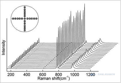
The data has three indicators: the position of the peak, the height of the peak (light intensity), and the width of the peak. Only when the peak position is completely matched, can it be 4H-SiC đủ điều kiện. Như với XRD, bất cứ khi nào có các đỉnh khác, chúng là các chất của các pha khác, đó là một khuyết tật.
The difference in the position of the peak is due to the difference in the energy of the phonons brought about by the different silicon carbide crystal lattices, that is, the different frequencies. Each phonon has its corresponding energy level. The virtual energy level theory can be used to explain Raman (non-linear process):
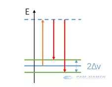
Hạt hấp thụ ánh sáng tới đến mức năng lượng ảo (màu cam), và sau đó chuyển trở lại mức năng lượng dao động (màu đỏ) khác với mức năng lượng ban đầu. Vì mức năng lượng trên là mức năng lượng ảo nên tần số của ánh sáng tới có thể thay đổi, miễn là nó không mâu thuẫn với mức năng lượng thực ban đầu.
Cần lưu ý rằng chế độ LOPC (964,769 cm-1) có thể được sử dụng để phân tích nồng độ chất mang:
n = 1.25 * 1017cm-2 *(964.769cm-1-VLOPC measurement)
As the carrier concentration increases, the interaction between atoms and the lattice increases, which makes the Raman peak blue shift (smaller), the intensity decreases, and the width increases. This method is not as accurate as other methods and can only be used as an aid to analyze the silicon carbide crystal distribution.
3. Why not Use XRD to Measure Silicon Carbide Crystal Distribution?
X-rays are optical radiation generated by the transition of electrons in the inner layer of atoms under the bombardment of high-speed moving electrons, including continuous X-rays and characteristic X-rays. Silicon carbide single crystal can be used as X-ray gratings, and the coherent scattering produced by these large numbers of particles (atoms, ions, or molecules) will cause light interference, increasing or decreasing the intensity of scattered X-rays. Due to the superposition of scattered waves from a large number of particles, the beams that interfere with each other to produce the highest intensity are called X-ray diffraction lines.
Để đáp ứng các điều kiện nhiễu xạ, có thể áp dụng công thức Bragg: 2dsinθ = nλ.
Chùm tia tới làm cho mỗi chất tán xạ bức xạ lại một phần nhỏ cường độ của nó như một sóng hình cầu. Nếu các chất tán xạ được sắp xếp đối xứng với nhau trong khoảng d, các sóng hình cầu này sẽ chỉ được đồng bộ theo hướng mà độ dài đường đi của chúng chênh lệch 2dsinθ bằng bội số nguyên của bước sóng λ. Trong trường hợp này, một phần của chùm tia tới bị lệch đi một góc 2θ, sẽ tạo ra các điểm phản xạ trong hình ảnh nhiễu xạ.
Use X-rays of known wavelengths to measure the θ angle to calculate the interplanar spacing d, which is used for X-ray structure analysis; the other is to use a silicon carbide seed crystal with a known d to measure the θ angle to calculate the characteristic X-ray wavelength, and then the elements contained in the sample can be found in the existing data.
The measurement formula is 2dSinθ=λ. While the d value among the cubic silicon carbide crystal is close, and the characteristic is not obvious enough, the accurate silicon carbide crystal distribution cannot be precisely determined. For these reasons, it is not suitable to use the XRD to measure the distribution of silicon carbide crystals.

Để biết thêm thông tin, vui lòng liên hệ với chúng tôi qua email victorchan@powerwaywafer.com và powerwaymaterial@gmail.com.